Islet Culture
Islet Culture SOPs: Islet culture, Determination of islet equivalents, Imaging islet samples, Digital imaging analysis of islet sample images
Islet Culture procedures
Flasks from the purification procedure are placed directly onto an inverted microscope for visual inspection. The trained islet personnel have learned to see islets without staining and to differentiate pure from imure fractions. Despite this training some fractions still need to be stained with dithizone (DMSO-free protocol published here) for determination of purity. Similar purities are combined and cultured together.
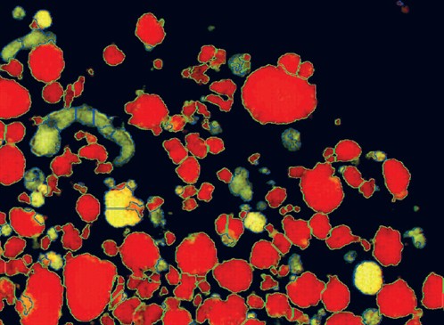
Islet samples are imaged and anaylzed using a digital image analysis system developed here in Uppsala. Image analysis reduces variability of both purity and islet counts as well as provides documentation of islet appearance for regulatory purposes.
 |
|
Figure: Digitally analyzed image of islets and exocrine
tissue |
Semi-closed culture bags
Our center developed the use of culture bags for islets whereas most others currently use flasks. The gas-permeable plastic allows for oxygen to diffuse directly to the islets while allowing a much larger volume of culture media to be used in comparison to flask-based culture methods. Islets remain metabolically active for weeks in this system.